Home » Uncategories » Sketch And Label Of A Cross Section Of A Long Bone / Skeletal System Labeled Diagrams Of The Human Skeleton - Some, mostly older, compact bone is remodelled to form these haversian systems (or osteons).the osteocytes sit in their lacunae in concentric rings around a central haversian canal (which runs longitudinally).the osteocytes are arranged in concentric rings of bone matrix called lamellae (little plates), and their processes run in interconnecting canaliculi.
Monday, 14 June 2021
Sketch And Label Of A Cross Section Of A Long Bone / Skeletal System Labeled Diagrams Of The Human Skeleton - Some, mostly older, compact bone is remodelled to form these haversian systems (or osteons).the osteocytes sit in their lacunae in concentric rings around a central haversian canal (which runs longitudinally).the osteocytes are arranged in concentric rings of bone matrix called lamellae (little plates), and their processes run in interconnecting canaliculi.
Sketch And Label Of A Cross Section Of A Long Bone / Skeletal System Labeled Diagrams Of The Human Skeleton - Some, mostly older, compact bone is remodelled to form these haversian systems (or osteons).the osteocytes sit in their lacunae in concentric rings around a central haversian canal (which runs longitudinally).the osteocytes are arranged in concentric rings of bone matrix called lamellae (little plates), and their processes run in interconnecting canaliculi.. Cross section = transverse section. In these labeled examples, a human femur is represented without identifying many of the unique characteristics that help differentiate the femur bone from other bones in the human body. A cross section of a compact bone shows concentric circles called lamellae. Label the haversian canal, osteocyte (mature bone cell) in lacuna, and canaliculi. Also known as the middle phalanx, the short pastern bone sits on top of the articulating joint of the pedal bone and underneath the long pastern bone.
Long bones have a thick outside layer of compact bone and an inner medullary cavity containing bone marrow. On this page, you will find two images i created that illustrate the parts of a long bone and long bone structure. Bone test anatomy and physiology 12 photos of the bone test anatomy and physiology anatomy and physiology bone lab test, anatomy and physiology bone markings test, anatomy and physiology bone practical test, anatomy and physiology bone tissue test, anatomy and physiology test on bone tissue, bone, anatomy and. Once we stop growing (between 18. The structure of a long bone allows for the best visualization of all of the parts of a bone (figure 6.7).
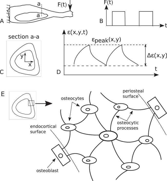
An Invertible Mathematical Model Of Cortical Bone S Adaptation To Mechanical Loading Scientific Reports from media.springernature.com Bone not color the articular cartilage; The diaphysis and the epiphysis. Sketch a longitudinal section through a long bone and label the following structures de epiphysim ercavi periosteum, co p pseen, compact bune.no red bone marrow, and yellow bone marrow he provides a epiphysis riedullary activity 4: The hollow region in the diaphysis is called the medullary cavity, which is filled. Osteon, central canal, blood vessels, lamellae, osteocytes, lacunae, canaliculi, and perforating canal. Make sure learners follow all the criteria for a biological drawing. The diaphysis and the epiphysis. Label the membrane that lines the cavity and the membrane that covers the outside surface.
Area between the diaphysis and epiphysis at both ends of the bone.
Label lines should not cross ; The hollow region in the diaphysis is called the medullary cavity, which is filled. A long bone has two parts: The diaphysis and the epiphysis. Fibrous layer (with fibroblasts) cellular layer ( chondroblasts). Osteon, central canal, blood vessels, lamellae, osteocytes, lacunae, canaliculi and perforating canal. • learn about the materials that make up bone • label a cross section of bone materials: Use colored pencils to draw and label the following structures as they appear using the 40x objective, or by looking at an image from the internet. The outside of a bone is covered in a thin layer of dense irregular connective tissue called the periosteum. Make sure learners follow all the criteria for a biological drawing. Draw a cross section of compact bone (microscopic view). The walls of the diaphysis are composed of dense and hard compact bone. Observed 2.sketch and label the diaphysis of the beef bone.
The ends of a long bone contain spongy bone and an epiphyseal line. The hollow region in the diaphysis is called the medullary cavity, which is filled with yellow marrow. Forms the larger rounded ends of long bones. The periosteum contains many strong collagen fibers that are used to firmly anchor tendons and muscles to the bone for movement. • computer with internet access • dr.
Structures Of The Ear In Chapter 04 Senses From Psychology An Introduction By Russ Dewey Ear Anatomy Human Ear Diagram Human Ear from i.pinimg.com Explain the functions of each of the labeled structures. This is for two reasons: Marks should be deducted for shading or colouring. Osteon, central canal, blood vessels, lamellae, osteocytes, lacunae, canaliculi, and perforating canal. The ends of a long bone contain spongy bone and an epiphyseal line. Anatomy of shoulder 12 photos of the anatomy of shoulder anatomy of nerves in shoulder, anatomy of posterior shoulder dislocation, anatomy of right shoulder, anatomy of shoulder labrum tear, anatomy of the shoulder games, human anatomy, anatomy of nerves in shoulder, anatomy of posterior shoulder dislocation, anatomy of. We start our section on tissue structure function with bone tissue. Use colored pencils to draw and label the following structures as they appear using the 40x objective, or by looking at an image from the internet.
Explain the functions of each of the labeled structures.
Label the height of the windows, either from the floor or ceiling. Once we stop growing (between 18. • computer with internet access • dr. In these labeled examples, a human femur is represented without identifying many of the unique characteristics that help differentiate the femur bone from other bones in the human body. Draw a cross section of compact bone (microscopic view). Label the haversian canal, osteocyte (mature bone cell) in lacuna, and canaliculi. External circumferential lamellae, osteon, central canal, perforating canals, lacuna, canaliculi, concentric lamellae. Make sure learners follow all the criteria for a biological drawing. Only the bottom portion of this bone extends as far as the hoof capsule. Label the membrane that lines the cavity and the membrane that covers the outside surface. Cow and human long bones have a similar general structure. Observed 2.sketch and label the diaphysis of the beef bone. Some, mostly older, compact bone is remodelled to form these haversian systems (or osteons).the osteocytes sit in their lacunae in concentric rings around a central haversian canal (which runs longitudinally).the osteocytes are arranged in concentric rings of bone matrix called lamellae (little plates), and their processes run in interconnecting canaliculi.
A long bone has two parts: Some, mostly older, compact bone is remodelled to form these haversian systems (or osteons).the osteocytes sit in their lacunae in concentric rings around a central haversian canal (which runs longitudinally).the osteocytes are arranged in concentric rings of bone matrix called lamellae (little plates), and their processes run in interconnecting canaliculi. Cow and human long bones have a similar general structure. This is the long central shaft. Label the haversian canal, osteocyte (mature bone cell) in lacuna, and canaliculi.
Cross Section Of The Mid Thigh Gross Anatomy Flashcards Draw It To Know It from d1j63owfs0b5j3.cloudfront.net Forms the larger rounded ends of long bones. Cross section = transverse section. Exploring the microscopic anatomy of bone 1. The diaphysis is the tubular shaft that runs between the proximal and distal ends of the bone. The periosteum contains many strong collagen fibers that are used to firmly anchor tendons and muscles to the bone for movement. A cross section of a compact bone shows concentric circles called lamellae. • computer with internet access • dr. Create a drawing of the bone section in your laboratory journal and label the areas listed above.
• computer with internet access • dr.
Draw a cross section of compact bone (microscopic view). Sketch a longitudinal section through a long bone and label the following structures de epiphysim ercavi periosteum, co p pseen, compact bune.no red bone marrow, and yellow bone marrow he provides a epiphysis riedullary activity 4: Volcano cross section diagram drawing high. Human left hand bone parts names. The ends of a long bone contain spongy bone and an epiphyseal line. Bone test anatomy and physiology 12 photos of the bone test anatomy and physiology anatomy and physiology bone lab test, anatomy and physiology bone markings test, anatomy and physiology bone practical test, anatomy and physiology bone tissue test, anatomy and physiology test on bone tissue, bone, anatomy and. The digital cushion sits just behind the pedal bone and above the sensitive frog. We start our section on tissue structure function with bone tissue. Draw and label a longitudinal section of a long bone. The diaphysis is the tubular shaft that runs between the proximal and distal ends of the bone. The diaphysis is the tubular shaft that runs between the proximal and distal ends of the bone. Use the internet or a reference textbook to help you identify the external features of long bone listed below. Anatomy of shoulder 12 photos of the anatomy of shoulder anatomy of nerves in shoulder, anatomy of posterior shoulder dislocation, anatomy of right shoulder, anatomy of shoulder labrum tear, anatomy of the shoulder games, human anatomy, anatomy of nerves in shoulder, anatomy of posterior shoulder dislocation, anatomy of.
0 Response to "Sketch And Label Of A Cross Section Of A Long Bone / Skeletal System Labeled Diagrams Of The Human Skeleton - Some, mostly older, compact bone is remodelled to form these haversian systems (or osteons).the osteocytes sit in their lacunae in concentric rings around a central haversian canal (which runs longitudinally).the osteocytes are arranged in concentric rings of bone matrix called lamellae (little plates), and their processes run in interconnecting canaliculi."


0 Response to "Sketch And Label Of A Cross Section Of A Long Bone / Skeletal System Labeled Diagrams Of The Human Skeleton - Some, mostly older, compact bone is remodelled to form these haversian systems (or osteons).the osteocytes sit in their lacunae in concentric rings around a central haversian canal (which runs longitudinally).the osteocytes are arranged in concentric rings of bone matrix called lamellae (little plates), and their processes run in interconnecting canaliculi."
Post a Comment